What is Transcranial direct current stimulation (tDCS)?

Overview
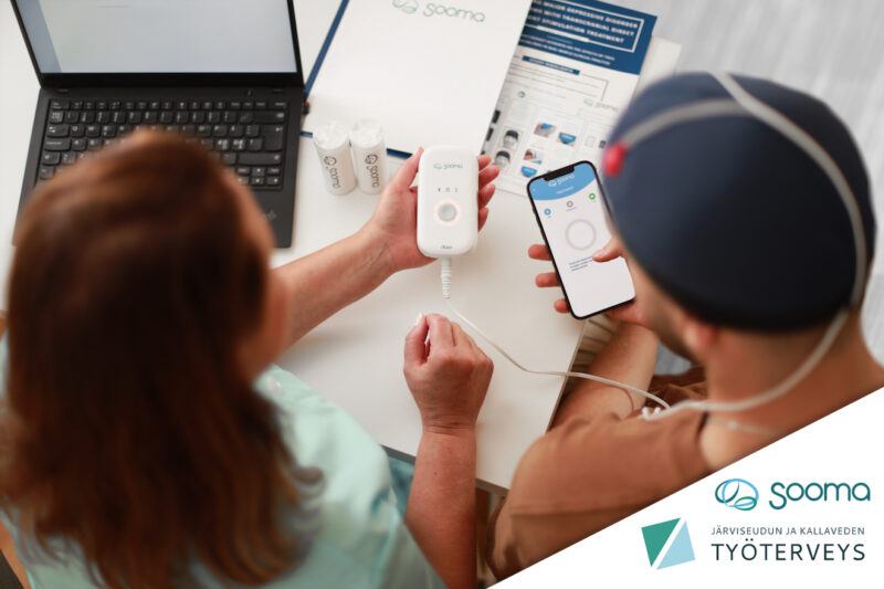
Transcranial direct current stimulation (tDCS) is a noninvasive neuromodulation technique used to treat a number of psychiatric and neurophysiological conditions. It uses a weak electric current, delivered to the brain from a usually small, portable battery-driven device. The stimulation area depends on what condition is being treated.
What is tDCS?
TDCS as a technique has its origins in early experimental studies on electricity with both animals and humans. In the 1960s, studies were conducted specifically on the neuronal effects of applying electricity to the brain, and it was discovered that mild electrical stimulation could modulate brain function by changing cortical excitability. The technique was named tDCS (transcranial direct current stimulation) in the 1990s, and for the past three decades, many studies have been conducted where tDCS has been applied to various psychiatric and neurophysiological conditions, such as depression, anxiety, schizophrenia, OCD, addiction, and different types of pain.
In tDCS, a weak current is used to stimulate targeted areas of the brain. The strength of the current is usually between 1 to 3 milliampere, with 2 milliampere being the most common when the treatment is clinically applied. The current travels from a small, portable battery-driven device to the brain via two electrodes; one anode, which is positively charged, and one cathode, which is negatively charged. The electrodes are positioned on the head using the 10-20 system commonly used in Electroencephalography (EEG) monitoring.
A treatment session usually lasts for 20 minutes, but can last up to 40 minutes. Sessions are often repeated to achieve a cumulative treatment effect. When behavioural changes are targeted, the session is repeated daily for several weeks or even longer in treatment protocols that aim to maintain a current condition. Aside from a short ramp-up and ramp-down at the start and end of the treatment, the current stays level throughout the treatment session.
What happens in the brain?
A human brain contains close to 100 billion neurons, which transmit electrical signals between each other to communicate. These electric signals are called action potentials. Action potentials are released, or “fired”, by neurons when there is a change in the neuronal resting membrane potential, which is the difference in electrical potential between a neuron’s inside and outside environment. In tDCS, the low current applied to the brain creates an electric field that polarises the resting membrane potential in the neurons of a targeted area. Typically, the resting membrane potential is made less negative, (“depolarised”) through excitatory stimulation in one area, and more negative (“hyperpolarised”) through inhibitory stimulation in another area. Depolarisation makes the neuron more likely to fire action potentials, increasing activity, while hyperpolarisation makes the neuron less likely to fire action potentials, decreasing activity. In tDCS, the stimulation does not force neurons to fire action potentials, but rather increases or decreases the likelihood of it by changing the amount of stimulus needed for the neuron to fire an action potential. The polarisation direction (depolarisation or hyperpolarisation) on a single neuron state depends on the neuron’s orientation relative to the electric field generated by tDCS.
How can we see the effects?
The fact that brief polarising currents can cause long-lasting after-effects on a rat brain was shown in-vivo already in the 1960s (Bindman et al, 1964). The polarising current influences neuronal excitability and modulates spontaneous neuronal firing rate during and after stimulation.
In humans, this effect has been demonstrated both directly and indirectly:
- MEP: The motor evoked potential amplitude is modulated by tDCS both during and after stimulation (Nitsche & Paulus, 2000)
- TMS-EEG: Global and local level changes in cortical excitability detected during and after stimulation (Romero-Lauro et al, 2014)
- MEG: Bilateral stimulation increases global connectivity and shows location and polarity dependent modulation of band power. (Hanley et al, 2016; Garcia-Cossio et al, 2016; Pellegrino et al, 2018)
- fMRI: Resting-state functional connectivity modulated by tDCS (Keeser et al, 2011)
- Implanted SCS: Amplitude changes in corticospinal pathway caused by tDCS in both I- and D-waves. (Lang et al, 2011; Di Lazzaro, 2013)
- Intracranial EEG: Electric fields of 0.8 V/m in the cortex during 2 mA stimulation (Huang et al, 2017)
- Implanted DBS: Voltage changes in the deep nuclei caused by tDCS stimulation (Chhatbar et al, 2018)
The immediate effects of tDCS are associated with changes in membrane potentials through voltage-dependent ion-channels. These can be diminished by blocking calcium- or sodium channels (Nitsche et al, 2003). The after-effects, while also reliant on membrane polarisation, are shown to be associated with NMDA-receptor function and the concentration of GABA neurotransmitters (Nitsche et al, 2006; Stagg et al, 2009). These effects translate into behavioural changes over repeated stimulation through functional connectivity and neuroplasticity (Jackson et al, 2016). The changes in neuroplasticity caused by tDCS are reported to involve several neurotransmitters such as dopamine, serotonin and acetylcholine. (Nitsche et al, 2006; Monte Silva et al, 2009; Kuo et al, 2007)
In depression

In depression, the left Dorsolateral Prefrontal Cortex (DLPFC) is known to be hypoactive (less active than normal) and the right Dorsolateral Prefrontal Cortex (DLPFC) is known to be hyperactive (more active than normal). To treat this imbalance in depression, a positively charged electrode (called an “anode”) is used to stimulate the left DLPFC with excitatory stimulation, and a negatively charged electrode (called a “cathode”) is used to stimulate the right DLPFC with inhibitory stimulation. This increases the likelihood of activity in the left DLPFC and decreases the likelihood of activity in the right DLPFC, eventually reaching the healthy balance found in individuals not suffering from depression.
In pain
In pain, tDCS is used to target the brain areas responsible for processing pain signals. The neurons in these areas use action potentials to communicate with each other and form neural circuits. According to Fregni et al (2020), recurring pain causes maladaptive neuroplasticity, which leads to a persistent sensation of pain, known as chronic pain. TDCS enables modulation of the maladaptive neuroplasticity and neuronal networks. Positive stimulation of areas C3 or C4, located in the primary motor cortex (M1), has been shown to reduce pain by enhancing the activity in these neural circuits. Excitatory stimulation is applied to M1 with the anode positioned on the opposite side of the pain (C3 or C4). A cathode is placed over the contralateral supraorbital area (O1 or O2, opposite side to the anode) to form a desirable electric field over the area being stimulated (M1). According to Lefaucheur et al (2017), excitatory stimulation to M1 enhances activity in the precentral gyrus, where the neuronal circuits connect to structures that are involved in the sensory and emotional parts of processing pain.
Latest news

TGA approves Sooma’s at-home brain stimulation for depression in Australia
Read more
Sooma Medical Announces Pivotal FDA IDE Clinical Trial for At-Home Brain Stimulation Device for Depression Treatment
Read more
Sooma Pioneers the Integration of Brain Stimulation into Primary Care, Improving Access to Early-Stage Depression Treatment
Read more